Ever wondered how a simple ECG can reveal the secrets of your heart? It all comes down to the leads on ECG—those vital connections that capture your heart’s electrical activity with precision and clarity.
What Are Leads on ECG and Why They Matter

The term leads on ecg refers to the specific views or perspectives of the heart’s electrical activity recorded by an electrocardiogram machine. These leads are not physical wires but rather combinations of electrodes placed on the body that measure voltage differences over time. Understanding these leads is crucial for accurate diagnosis of cardiac conditions such as arrhythmias, myocardial infarctions, and conduction abnormalities.
Definition of ECG Leads
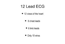
In electrocardiography, a ‘lead’ represents a particular electrical pathway between two or more electrodes. Each lead provides a unique angle from which the heart’s depolarization and repolarization can be observed. The standard 12-lead ECG uses 10 electrodes to generate 12 different views—4 limb leads and 8 precordial (chest) leads.
- Leads are mathematical derivations of electrode voltages.
- They reflect the direction and magnitude of electrical impulses in the heart.
- Each lead corresponds to a specific anatomical region of the heart.
Types of Leads: Limb vs. Precordial
There are two primary categories of leads on ecg: limb leads and precordial (chest) leads. Limb leads are placed on the arms and legs and include both standard bipolar leads (I, II, III) and augmented unipolar leads (aVR, aVL, aVF). Precordial leads (V1–V6) are positioned across the chest wall to provide horizontal plane views of the heart.
- Limb leads assess the heart in the frontal plane.
- Precordial leads evaluate the heart in the horizontal (transverse) plane.
- Together, they create a 3D representation of cardiac electrical activity.
“The 12-lead ECG is one of the most powerful diagnostic tools in cardiology—when interpreted correctly, it can detect life-threatening conditions within seconds.” — Dr. Eric Topol, Scripps Research
How Many Leads Are There on a Standard ECG?
A standard clinical ECG consists of 12 leads on ecg, derived from 10 electrodes attached to the patient’s skin. Despite the name, these 12 leads do not mean 12 separate wires; instead, they are calculated using combinations of electrode inputs. This system allows clinicians to view the heart from multiple angles, increasing diagnostic accuracy.
The 12-Lead Configuration Explained
The 12-lead ECG includes six limb leads (I, II, III, aVR, aVL, aVF) and six precordial leads (V1 to V6). The limb leads are further divided into two groups: the three standard limb leads (I, II, III), which are bipolar, and the three augmented limb leads (aVR, aVL, aVF), which are unipolar. The precordial leads are all unipolar and placed across the chest at specific intercostal spaces.
- Lead I: Right arm to left arm
- Lead II: Right arm to left leg
- Lead III: Left arm to left leg
- aVR: Augmented vector right
- aVL: Augmented vector left
- aVF: Augmented vector foot
- V1–V6: Chest electrodes from 4th intercostal space right sternal border to midaxillary line
Why 12 Leads? The Diagnostic Advantage
The use of 12 distinct leads on ecg enables comprehensive spatial coverage of the heart. This multi-angle assessment helps localize ischemic changes, identify infarct locations, and detect chamber enlargement. For example, ST-segment elevation in leads II, III, and aVF suggests an inferior wall myocardial infarction, while changes in V1–V3 point to anterior wall involvement.
- Provides full 360-degree electrical mapping of the heart.
- Enables localization of myocardial damage.
- Facilitates early detection of life-threatening arrhythmias.
For more detailed information on ECG lead configurations, visit the Healio ECG Review, a trusted resource for medical professionals.
Understanding Limb Leads on ECG
The limb leads on ECG are essential for interpreting the heart’s electrical axis in the frontal plane. These six leads—three standard and three augmented—form the foundation of ECG interpretation and are critical for identifying rhythm disturbances and axis deviations.
Standard Bipolar Limb Leads (I, II, III)
These leads measure the voltage difference between two limbs. Lead I compares the left arm to the right arm, Lead II the left leg to the right arm, and Lead III the left leg to the left arm. Together, they form Einthoven’s Triangle, a conceptual model that underpins much of ECG theory.
- Lead I: Positive on left arm, negative on right arm
- Lead II: Positive on left leg, negative on right arm
- Lead III: Positive on left leg, negative on left arm
Augmented Unipolar Limb Leads (aVR, aVL, aVF)
Unlike the bipolar leads, aVR, aVL, and aVF are unipolar, meaning they measure the voltage at one electrode relative to a combined reference point from the other two. These leads enhance sensitivity and provide additional perspectives:
leads on ecg – Leads on ecg menjadi aspek penting yang dibahas di sini.
- aVR: Looks at the heart from the right shoulder
- aVL: Views the lateral left side
- aVF: Sees the inferior surface
“aVR is often overlooked, but it can be a key indicator in global ischemia or dextrocardia.” — Dr. Amal Mattu, Emergency Cardiology Expert
The Role of Precordial Leads on ECG
Precordial leads, also known as chest leads, are central to analyzing the horizontal plane of the heart. These six leads—V1 through V6—are placed directly on the chest and offer detailed insight into the anterior, septal, and lateral walls of the left ventricle.
Placement of Chest Leads (V1–V6)
Correct placement of precordial electrodes is vital for accurate readings. Misplacement can lead to misdiagnosis. Here’s the standard positioning:
- V1: 4th intercostal space, right sternal border
- V2: 4th intercostal space, left sternal border
- V3: Midway between V2 and V4
- V4: 5th intercostal space, midclavicular line
- V5: Same horizontal level as V4, anterior axillary line
- V6: Same level as V4, midaxillary line
For visual guidance on proper placement, refer to the ECG Waves educational site.
Clinical Significance of Precordial Leads
Precordial leads are indispensable in diagnosing anterior myocardial infarctions, bundle branch blocks, and ventricular hypertrophy. For instance:
- ST elevation in V1–V4 suggests anterior MI
- Deep S waves in V1 and tall R waves in V5–V6 indicate left ventricular hypertrophy
- Wide, notched R waves in V1 may point to right bundle branch block
How Leads on ECG Map the Heart’s Electrical Activity
Each of the leads on ecg corresponds to a specific anatomical region of the heart. By analyzing which leads show abnormalities, clinicians can pinpoint the location of cardiac pathology with remarkable accuracy.
Correlation Between Leads and Heart Walls
Understanding which lead views which part of the heart is fundamental in ECG interpretation:
- Inferior wall: Leads II, III, aVF
- Lateral wall: Leads I, aVL, V5, V6
- Anterior wall: V3, V4
- Septal wall: V1, V2
- Right ventricle: V1, V2 (and sometimes V4R for right-sided infarcts)
- Posterior wall: Indirectly assessed via V1, V2 (tall R waves, ST depression)
Electrical Axis Determination Using Leads
The QRS axis in the frontal plane is determined primarily by examining the net deflection in leads I and aVF. A normal axis ranges from -30° to +90°. Deviations can indicate conditions like left or right axis deviation, often due to ventricular hypertrophy or conduction blocks.
- Positive in I and aVF = normal axis
- Negative in I, positive in aVF = right axis deviation
- Positive in I, negative in aVF = left axis deviation
- Negative in both = extreme axis deviation (rare)
“Axis determination is the first step in systematic ECG interpretation—it sets the stage for everything else.” — Life in the Fast Lane Medical Education
Common Errors in Lead Placement and Their Impact
Misplaced electrodes are a frequent source of ECG misinterpretation. Even small deviations in leads on ecg placement can mimic pathology or mask real disease.
Effects of Incorrect Limb Lead Placement
Swapping limb electrodes can drastically alter the appearance of the ECG. For example:
- Right and left arm reversal: Inverts lead I and causes confusion in axis calculation
- Arm-leg swap: Can mimic dextrocardia or limb lead reversal patterns
- Reversed leg leads: Usually less impactful but can distort inferior leads
Consequences of Precordial Lead Misplacement
Placing chest leads too high or too low can simulate or obscure myocardial infarction patterns. For instance:
- V1 and V2 placed too high may show false ST elevation
- V4–V6 misplaced posteriorly can mimic lateral ischemia
- Failure to place V4 at the 5th ICS midclavicular line reduces sensitivity for anterior MI
A study published in the National Center for Biotechnology Information (NCBI) found that up to 40% of ECGs have some degree of lead misplacement, emphasizing the need for standardized training.
leads on ecg – Leads on ecg menjadi aspek penting yang dibahas di sini.
Advanced Applications of Leads on ECG
Beyond the standard 12-lead ECG, advanced applications leverage the principles of leads on ecg to improve diagnostic precision in complex cardiac cases.
Right-Sided ECG (V4R)
In suspected right ventricular infarction (often accompanying inferior MI), placing a right-sided lead V4R (same position as V4 but on the right side) can detect ST elevation, guiding fluid management and thrombolytic therapy.
- V4R: 5th intercostal space, right midclavicular line
- ST elevation in V4R has high specificity for right ventricular involvement
- Used in conjunction with standard 12-lead ECG
Posterior Leads (V7, V8, V9)
When posterior myocardial infarction is suspected (e.g., tall R waves in V1–V2), posterior leads are placed:
- V7: Left posterior axillary line, same level as V6
- V8: Left scapular line, same level
- V9: Left paraspinal area, same level
- ST elevation in these leads confirms posterior MI
Esophageal and Intracardiac Leads
In electrophysiology studies, specialized leads are inserted into the esophagus or directly into the heart chambers to record electrical activity with greater resolution. These are used to diagnose and ablate complex arrhythmias like atrial flutter or WPW syndrome.
- Esophageal leads: Close proximity to the atria enhances P-wave visibility
- Intracardiac leads: Used during catheter ablation procedures
- Provide real-time, high-fidelity data not possible with surface ECG
Interpreting Abnormalities Across Leads on ECG
Mastering the interpretation of leads on ecg involves recognizing patterns across multiple leads. No single lead tells the whole story—context is everything.
Recognizing Myocardial Infarction Patterns
Acute MI produces characteristic changes in specific leads:
- Inferior MI: ST elevation in II, III, aVF
- Anterior MI: ST elevation in V1–V4
- Lateral MI: ST elevation in I, aVL, V5, V6
- Posterior MI: ST depression in V1–V3, confirmed with V7–V9
- Right ventricular MI: ST elevation in V4R
Identifying Arrhythmias Using Lead Patterns
Different arrhythmias manifest uniquely across leads:
- Atrial fibrillation: Irregularly irregular rhythm, absent P waves in multiple leads
- VT vs. SVT with aberrancy: Axis, QRS width, and AV dissociation clues in limb leads
- Brugada pattern: Coved ST elevation in V1–V3
- Long QT syndrome: Prolonged QT interval best measured in lead II or V5
“The ECG is a language. Each lead is a word, and together they form sentences that tell the story of the heart.” — Dr. Philip Podrid, Boston University School of Medicine
Training and Best Practices for ECG Lead Use
Proper training in leads on ecg placement and interpretation is essential for healthcare providers. Errors in technique can lead to misdiagnosis, delayed treatment, or unnecessary interventions.
Standardized Protocols for Lead Placement
Hospitals and clinics should adopt standardized protocols to ensure consistency:
- Use anatomical landmarks (e.g., angle of Louis, midclavicular line)
- Verify electrode contact and skin preparation
- Double-check lead wire connections
- Document any deviations from standard placement
Education and Simulation-Based Training
Simulation labs and online modules can improve competency in ECG interpretation. Resources like ECG Library offer case-based learning with real tracings.
- Interactive ECG simulators enhance pattern recognition
- Peer review of ECGs improves diagnostic accuracy
- Regular refresher courses maintain skill proficiency
For comprehensive training materials, visit the American College of Cardiology’s ECG resources.
leads on ecg – Leads on ecg menjadi aspek penting yang dibahas di sini.
What are the 12 leads on an ECG?
The 12 leads on an ECG consist of six limb leads (I, II, III, aVR, aVL, aVF) and six precordial leads (V1–V6). These leads provide a comprehensive view of the heart’s electrical activity from multiple angles, enabling accurate diagnosis of cardiac conditions.
What happens if ECG leads are placed incorrectly?
Incorrect lead placement can lead to misinterpretation of the ECG, potentially resulting in false diagnoses such as myocardial infarction or arrhythmia. For example, swapping arm electrodes can invert lead I and mimic dextrocardia, while misplaced chest leads can obscure ST-segment changes.
Can ECG leads detect a heart attack?
Yes, leads on ecg are critical in detecting heart attacks. Specific leads show ST-segment elevation or depression, T-wave inversions, or new Q waves that indicate myocardial injury. The location of these changes helps determine which part of the heart is affected.
What is the purpose of augmented limb leads?
Augmented limb leads (aVR, aVL, aVF) enhance the signal from unipolar electrodes by amplifying the voltage. They provide additional perspectives on the heart’s frontal plane activity and are especially useful in axis determination and identifying global ischemia.
How do posterior leads help in ECG diagnosis?
Posterior leads (V7, V8, V9) are placed on the back to detect posterior myocardial infarction, which may not be visible on standard leads. ST elevation in these leads confirms posterior wall involvement, often seen with tall R waves and ST depression in V1–V3.
Understanding leads on ecg is fundamental to mastering electrocardiography. From the precise placement of electrodes to the interpretation of complex waveforms, each lead plays a vital role in revealing the heart’s electrical story. Whether diagnosing a myocardial infarction, identifying arrhythmias, or detecting chamber enlargement, the 12-lead ECG remains an indispensable tool in modern medicine. By adhering to best practices in lead placement and interpretation, clinicians can ensure accurate, timely, and life-saving diagnoses.
leads on ecg – Leads on ecg menjadi aspek penting yang dibahas di sini.
Further Reading: